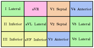
Axis
- -30 to 90 is normal
- Determine Axis
- Look at I, II, and aVL QRS
- I up ⇒ Right of vertical axis
- II up ⇒ Between -30 and 150
- aVF up ⇒ Below horizontal axis
| Axis Direction | Interpretation | |
|---|---|---|
| I: ↑ II: ↑↓ aVF: ↑ |
0 - 90 | Normal |
| I: ↑ II: ↑ aVF: ↓ |
-30 - 0 | Small LAD |
| I: ↑ II: ↓ aVF: ↓ |
-90 - -30 | LAD |
| I: ↓ II: ↑ aVF: ↑ |
90-150 | RAD |
| I: ↓ II: ↓ aVF: ↑ |
150 - 180 | RAD |
| I: ↓ II: ↓ aVF: ↓ |
-180 - -90 | Extreme Axis Deviation |
Rhythm Analysis1
- Normal Rate: 60-100
- Normal P-wave: < 0.12s
- Often biphasic in V1
- Slight notch in precordial leads is normal
- Notch w/ peak-peak interval > 1mm (0.04s) is usually pathological
- Normal PR Interval: 0.12-0.2s
- Normal QRS-complex: < 0.1s
- Non-pathological “septal” Q waves often present in I, aVL, V5, V6
- Septal Q waves should be < 2 small squares deep and < 25% of R-wave amplitude
- Non-pathological “septal” Q waves often present in I, aVL, V5, V6
- Normal QTc: < 0.5s
- Shortcut: If T wave is at or past halfway between the preceding and next QRS complex w/o tachycardia, it is likely prolonged
- QTc = $\text{QT}/\sqrt{\text{R-R interval (s)}}$
- Normal T Wave
- Inversion in V1 normal, sometimes in V2
AV Conduction Delays
- 1st Degree HB
- PR interval > 0.2s
- 2nd Degree HB
- Mobitz Type I (Wenckebach): PR progressively lengthens until QRS is dropped, then returns to normal
- Mobitz Type II: Intermittent failure, usually in a fixed ration, but PR interval does not change
- 3rd Degree HB
- No correlation between P and QRS, but both can still occur, QRS can be narrow if AV node is firing, or wide if ventricles are pacing
Bundle Branch Blocks
- RBBB
- QRS ≥ 0.12s
- R’ in V1 or V2 (looks like a big M)
- Slurred S wave in I, V5, and V6
- Optional: ST depression and T wave inversion in right precordial leads
- LBBB
- QRS > 0.12s
- Broad, monophonic R wave in I, V5, and V6
- No Q wave in V5 or V6
- Optional: ST-T displacement discordant to QRS complex
- Optional: Poor R-wave progression
- Optional: RS Complex instead of monophonic complex in V5 and V6
- Optional: Left Axis Deviation (common, but not required)
- Fascicular Block
- Anterior Block: Axis < -30
- Posterior Block: Axis > 90
- Bifasicular (RBBB and LA or LPBBB): RBBB and LABBB is more common, variable axis, but deviated
- Trifasicular Block: Bifasicular + 1st Degree block => Complete heart block
Bradycardias
- Sick Sinus Syndrome
- Rate < 60, treat only if SSx
- SA Block vs SA Arrest
- SA block: Pause length is a multiple of normal P-P interval
- SA Arrest: Pause is not a multiple of normal P-P interval
Atrial Tachycardias
- Sinus Tach
- Normal morphology and rate > 100, rarely exceeds 200 BPM
- Afib
- 350-600 BPM atrial rate
- 100-180 BPM ventricle rate
- Aflutter
- 300 BPM atrial rate, morphologically distinct, unlike afib
- Ventricles usually conducted 2:1, so rate is usually 150 BPM
- Atrial Tach
- Ectopic atrial pulses of 150-250 BPM, notably P waves are abnormal compared to sinus tach
- Usually 1:1 conduction
- Atrial tach w/ AV block is commonly seen w/ digoxin toxicity
Junctional Tachycardias
- Normal QRS morphology w/o accessory pathways (WPW)
- AVNRT
- Re-entry is in the node, leads to rapid ventricular rate with normal or absent P waves, and normal, regular QRS complexes
- P-waves can occur at the end of the QRS (backwards conduction), leading to pseudo-S waves or pseudo-R waves depending on lead
- AVRT / WPW
- Occur due to accessory pathway
- WPW is prototypical, and can be conducted “prodromic” (normal direction) or “antidromic” (backwards)
- Orthodromic divided into two types: A and B
- Type A: Large R wave in all leads, may be RR’ in V1, can resemble RBBB, RVH, or posterior MI
- Type B: Large S in V1-3 and r in V4-V6, can resemble LBBB and anterior MI
- Antidromic AVRTs have wide complexes, abnormal looking rhythm
- Afib and WPW Can resemble vtach, w/ irregular broad complexes and the occasional narrow complex when conducted appropriately
Wide-Complex Tachycardias
- Changes in axis +/- 40 degree when arrhythmia arises ⇒ ventricular arrhythmia
- Vtach vs SVT w/ BBB
- RBBB Morphology: Ventricular if QRS > 0.14s, Axis deviation, QS wave or negative complex in V6, concordance throughout chest leads, R or biphasic R in V1
- LBBB Morphology: Venctricular if axis deviation, QRS &gr; 0.16s, QD w/ negative complex in V6, concordance through chest leads, rS in V1
#Ischemia / Infarct
#Hypertrophy
Reference
-
ABCs of Clinical Electrophysiology. BMJ. Section 1-15. ↩